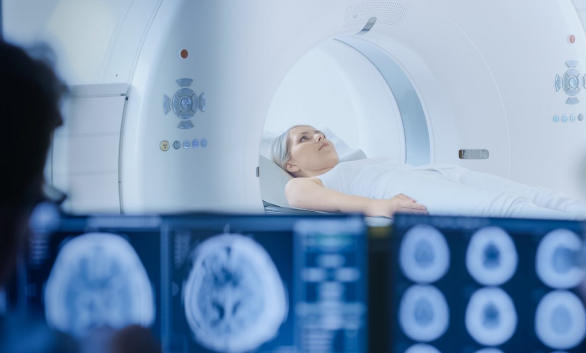
There are many blog posts about the positive results of Deep Brain Stimulation (DBS) surgery for targeted relief of dystonia symptoms. When considering this procedure, it is important to consider the role of MRI prior to and post-surgery in determining if this treatment is a suitable option for your lifestyle.
Magnetic Resonance Imaging (MRI) uses powerful magnets to produce three-dimensional anatomical images. In most MRI machines, the patient is placed inside a long tube where they lay very still while the technician pulses a series of radiofrequencies through the tube, resulting in images as the protons in the body realign due to the interaction between the radiofrequencies and the magnet. An MRI is completely safe for most patients, but there are exceptions.
- Due to the magnets involved, people with implants, especially those containing iron, should avoid an MRI. These implants include pacemakers, vagus nerve stimulators, implantable cardioverter-defibrillators, insulin pumps, cochlear implants, and some joint replacements. If you are unsure whether an implanted device contains metals that would interact with the magnets, notify your doctor or MRI technician.
- People who are particularly sensitive to noise or vibrations may have difficulty completing an MRI. The sound intensity in some MRI scanners can reach 120 decibels. Noise canceling headphones diminish the volume, but they do not completely remove the sound. The pulsing of the machine may also be difficult for patients who are particularly sensitive to tactile sensations.
- Individuals who are claustrophobic may have difficulty completing lengthy scans in traditional MRI machines. Relaxation/visualization techniques or using an open-sided MRI may be helpful in overcoming this barrier as sedation is typically not available for dystonia patients seeking an MRI for DBS surgery.
- An MRI is not recommended during the first trimester of pregnancy, though this recommendation is listed as a precaution. No specific harm to the developing fetus has been documented.
DBS patients may receive an MRI at three points in their surgical journey. Some physicians may request an MRI as part of the evaluation process to determine if a patient is a strong candidate for DBS. This evaluation process may include CT/MRI imaging, neuropsychiatric testing, review of previous treatments and their outcomes, and development of a recovery support plan. The second MRI may occur immediately prior to surgery if the surgeon is using the stereoaxis method. After receiving a light anesthetic, a frame is applied to the patient’s head. With the frame in place, the patient receives an MRI or CT scan to calculate the correct placement for the DBS leads. The patient then goes directly to surgery for placement of the leads. A third MRI or CT may occur immediately after surgery to confirm the leads have been placed correctly. There are currently investigational protocols in which the entire surgery is completed within a high-resolution MRI scanner.
Once the DBS leads are in place, a pulse generator unit is inserted in the patient’s chest. After this process is complete, the patient is no longer able to receive an MRI according to standard MRI protocols due to the level of metal in his/her body. Some MRI facilities would be willing to complete some MRI protocols on an individual with a DBS device using a “low energy” protocol. This depends on the specific device inserted, the duration of the protocol required, and the MRI machine being utilized.
As with all medical decisions, you should consult your doctor to determine if DBS is right for you. If you have concerns about how you would tolerate MRIs before or during the DBS surgery or the potential need for MRIs after DBS surgery, discuss them with your doctor when determining if this is the correct treatment option for you.
Sources:
National Institute of Biomedical Imaging and Bioengineering: Magnetic Resonance Imaging
